


パーキンソンと診断されてから通院を何度もしていました。ですが、こちらで購入してからは家から出なくてもいいので、今ではこちらのサイトでお世話になっています。通院の付き添いで家族に付き合ってもらうのも悪いですから、こういうサイトがあるのは非常に助かります。

左記クレジットカード、銀行振込、コンビニ決済に対応
左記クレジットカード、銀行振込、コンビニ決済に対応


更新日:2025/6/9

| 個数 | 販売価格(1錠あたり) | 販売価格(箱) | ポイント | 購入 |
|---|---|---|---|---|
| 8錠 | 812円 | 6,500円 | 195pt | |
| 16錠 | 650円 | 10,400円 | 312pt | |
| 24錠 | 583円 | 14,000円 | 420pt |


①1万円以上で送料無料
1回の注文で10,000円以上だった場合、1,000円の送料が無料となります。
まとめ買いをすると1商品あたりのコストパフォーマンスが高くなるためおすすめです。
②プライバシー守る安心梱包
外箱に当サイト名や商品名が記載されることはないため、ご家族や配達員など第三者に内容を知られることは御座いません。

③100%メーカー正規品取り扱い
当サイトの商品は100%メーカー正規品となっており、第三者機関による鑑定も行っております。
商品の破損などがあった場合は再配送などにて対応させて頂きますので、ご連絡頂ければ幸いです。

④いつでも購入可能 処方箋不要
サイト上では24時間いつでもご注文を受けております。
また、お電話によるご注文も受け付けておりますのでネットが苦手な方はお気軽にどうぞ。

⑤商品到着100%
商品発送後はお荷物の追跡状況が分かる追跡番号をご案内させて頂きます。
郵便局には保管期限がありますのでご注意ください。
・自宅配達で不在だった場合の保管期限・・・16日間前後
・郵便局留めとした場合の保管期限・・・7~30日間

⑥コンビニ決済利用可能
ご近所のコンビニにていつでもお支払可能です。
セブンイレブンに限り店舗での機械操作を必要とせず、手続き完了後に表示されるバーコードや払込票番号をレジに提示することでお支払い頂けます。

カベルゴリン 0.5mg x 8錠
6,500円
ポイント:195pt
10,000円以上購入で送料無料
在庫あり

パーキンソンと診断されてから通院を何度もしていました。ですが、こちらで購入してからは家から出なくてもいいので、今ではこちらのサイトでお世話になっています。通院の付き添いで家族に付き合ってもらうのも悪いですから、こういうサイトがあるのは非常に助かります。
飲み始めて数日。副作用がちょっと辛くて飲めないかも。自分にはあってなさそうだから、他の薬にしてみる。
薬が登場する前は、パーキンソン病の自覚症状があらわれてから5年程で歩行困難、7年目までには寝たきりとなるとされていました。そのため、パーキンソン病を自覚しているといったような場合には適切に治療することが重要です。
現在パーキンソン病の治療薬として使われているものは特効薬ではありません。病気の進行を遅らせる作用を持ったものしかなく、薬を一度飲んで完治させるといったことは不可能です。
コーヒーやお酒などを絶対に飲めなくなるということは考えにくいですが、何かしら別の理由で飲酒などが禁止されているケースもあります。そのため、医師に相談した上で飲酒などの許可が出れば問題ないと考えられます。
パーキンソン病の治療薬はあくまでも進行を遅らせるものでしかありません。そのため、完全に進行を止めるといったことはできません。ある程度の改善は見込めますが、徐々に悪化していくという点については把握しておく必要があります。
| 1日の服用回数 | 1回 |
|---|---|
| 1回の服用量 | 0.25~0.5mg |
| 服用のタイミング | 朝食後 |
| 服用間隔 | 24時間以上 |
| 1日の服用回数 | 1回 |
|---|---|
| 1回の服用量 | 0.25mg |
| 服用のタイミング | 就寝前 |
| 服用間隔 | 24時間以上 |
| 1日の服用回数 | 1回 |
|---|---|
| 1回の服用量 | 1mg |
| 服用のタイミング | 出産後の食後 |
| 服用間隔 | 1回のみ |
| 商品名 | ベンズトロップ | アマンタジン | オンジェンティス | PK-メルツ | コマントレル-T | サフィオン | レボドパ/カルビドパ/エンタカポン | タスマール | ロパーク |
|---|---|---|---|---|---|---|---|---|---|
| 商品画像 |  |  |  |  |  |  |  |  |  |
| 特徴1 | ・ドパミンの活性を増加 | ・抗インフルエンザウイルス作用にも期待できる | ・パーキンソンの治療薬として国内でも処方 | ・パーキンソン病の治療薬 | ・パーキンソン病だけでなくA型インフルエンザにも有効 | ・ドパミンの分解を抑制 | ・従来薬よりもさらに高い効果を期待できる | ・脳内のドパミン濃度を上昇 | ・効果が安定しているドパミン作動薬 |
| 特徴2 | ・パーキンソン病の症状を軽減 | ・副作用が少なく、他の医薬品との併用も可能 | ・パーキンソン病の症状を改善 | ・ドイツの有名メーカーが作るパーキンソン病治療薬で安心 | ・ドパミンの不足を効果的に解消できる | ・脳内のドパミン量が増加 | ・ドパミンの不足を補って症状を改善する | ・手足の震えや筋肉のこわばりを改善 | ・震えやこわばりを改善 |
| 内容量 | 2mg60錠x1本 | 100mgx30錠 | 50mgx10錠 | 100mg90錠x1箱 | 100mg100錠x1箱 | 50mg100錠x1箱 | 50mg+12.5mg+200mg 100錠x1箱 | 100mg100錠x1本 | 0.25mg100錠x1箱 |
| 価格 | 5,960円 | 4,360円 | 12,360円 | 8,760円 | 6,560円 | 6,560円 | 10,560円 | 25,360円 | 5,560円 |
本製品は海外製のため、期限表記が日本と異なる場合がございます。
パッケージ裏面や側面、シートなどに以下のような表記がされています。
| EXP | 使用期限 例:EXP 12/2025→2025年12月まで使用可 |
|---|---|
| MFG または MFD | 製造日 例:MFG 03/2023 |
| BEST BEFORE | 品質が最も安定している目安日 |
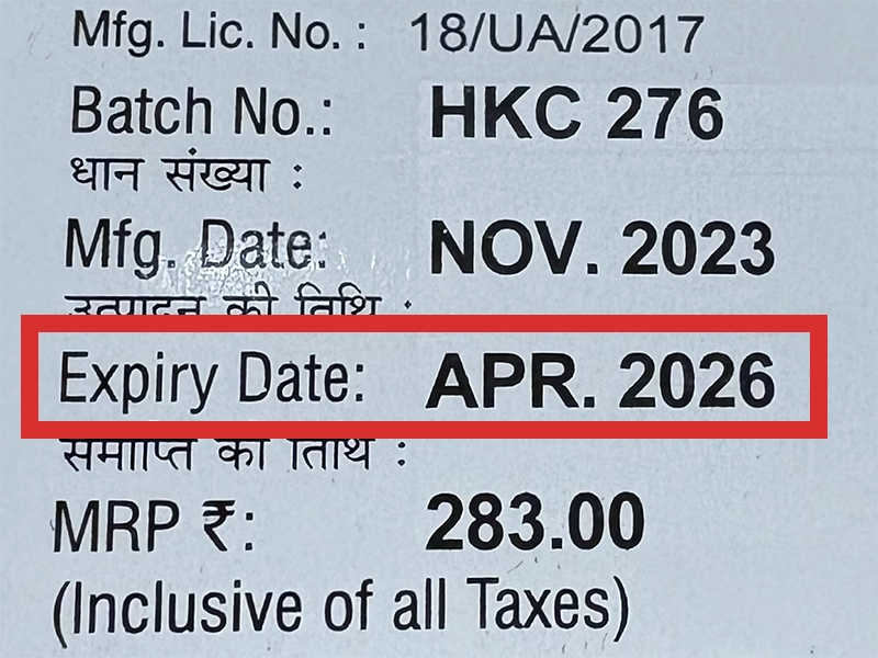
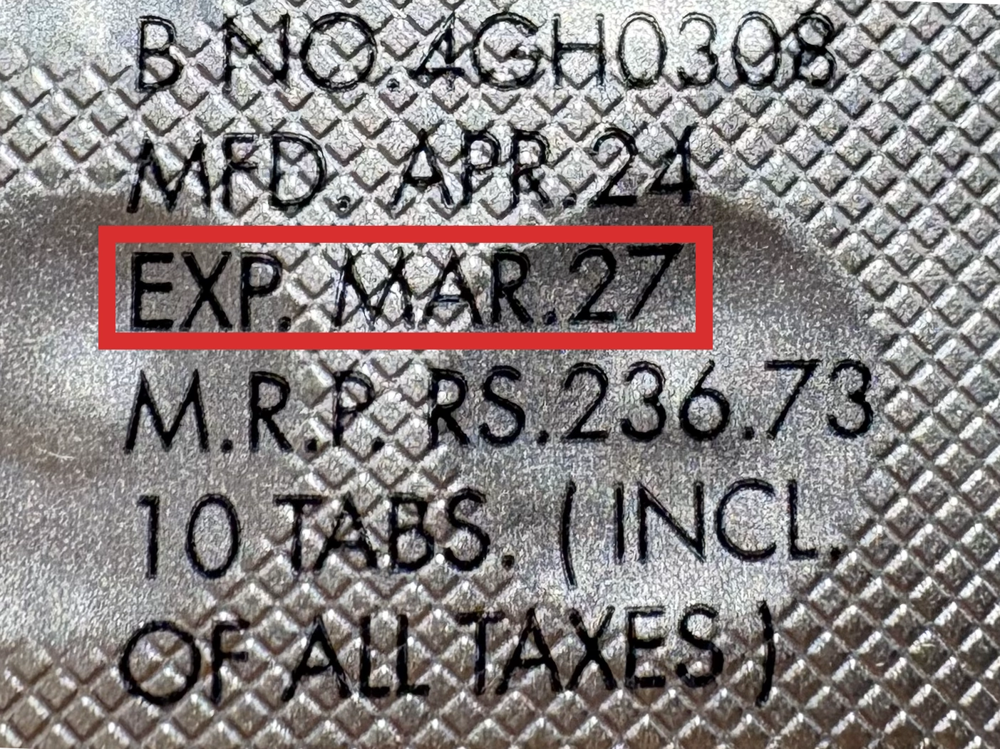
※国や製品により日付の並び(例:月/年、日/月/年)が異なる場合がありますのでご注意ください
EXP(Expiry Date) の表記がなく、MFG または MFDしか記載がないケースがあります。
この場合は MFG(MFD) から2~3年が使用期限の目安です。
※「LOT」や「BATCH」の表記は製造番号であり期限ではありません。

パッケージ例となります。
商品やご注文単位によってはシート単位でのお届けとなる場合が御座います。
外箱に当サイト名や商品名が記載されることはないため、ご家族や配達員など第三者に内容を知られることは御座いません。
パーキンソンと診断されてから通院を何度もしていました。ですが、こちらで購入してからは家から出なくてもいいので、今ではこちらのサイトでお世話になっています。通院の付き添いで家族に付き合ってもらうのも悪いですから、こういうサイトがあるのは非常に助かります。
改善までとはいきませんが、薬が効いている間は症状を少し抑えてくれます。震えがとまらないと、普通に生活するのにも大変ですからね。病院で処方された薬よりも副作用が気にならないので、私はこちらの薬が気に入っています。
飲み始めて数日。副作用がちょっと辛くて飲めないかも。自分にはあってなさそうだから、他の薬にしてみる。
処方箋なしでパーキンソン病の薬を購入できるのは凄いですね。通院するのが辛い私からすると、凄く助かります。届くまでには何週間かかかりますが、前もって注文しておけば薬を切らす事はないです。
意外と安くて驚きましたね。このぐらいの値段なら続けて購入するのもツラくはないかも。
商品口コミの投稿は会員のみ行えるようになっております。
お手数ですが会員ログインの上でご投稿頂きますようお願いいたします。
口コミをご投稿頂いたお客様にはポイントをプレゼントさせて頂いております。
文章のみであれば100ポイント、文章+写真付きのものは300ポイントをプレゼントさせて頂きます。
規約や詳細などはこちらをご確認くださいませ。